


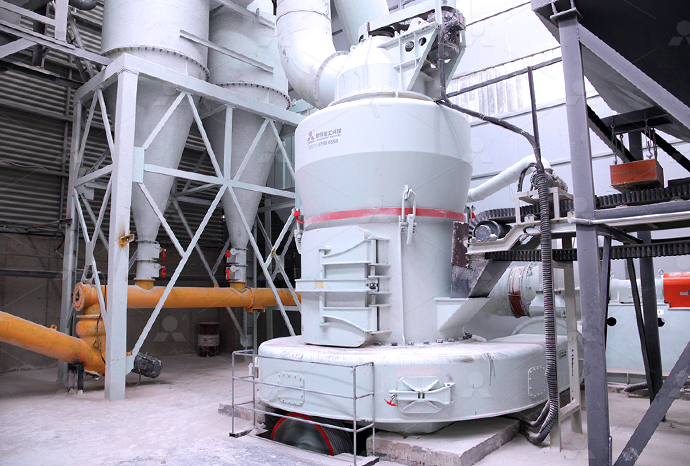
MTW European Type Trapezium Mill
Input size:30-50mm
Capacity: 3-50t/h
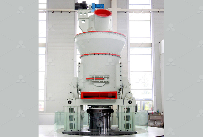
LM Vertical Roller Mill
Input size:38-65mm
Capacity: 13-70t/h
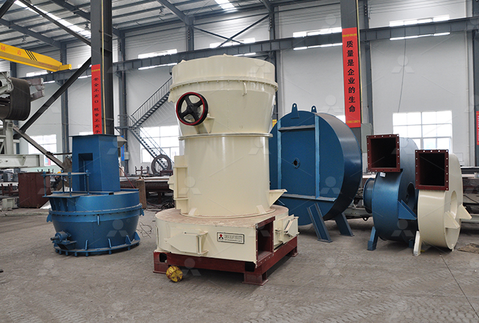
Raymond Mill
Input size:20-30mm
Capacity: 0.8-9.5t/h
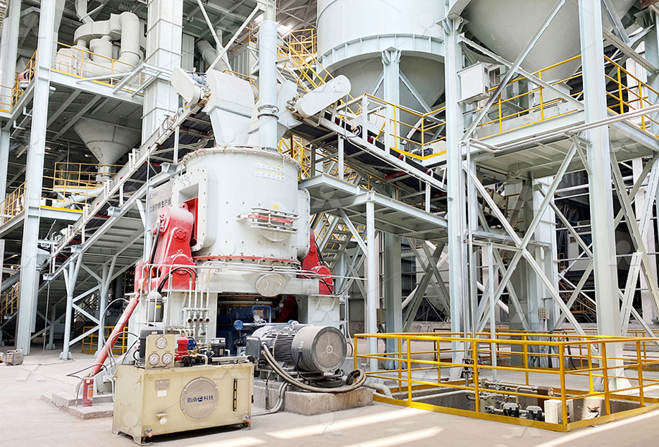
Sand powder vertical mill
Input size:30-55mm
Capacity: 30-900t/h
LUM series superfine vertical roller grinding mill
Input size:10-20mm
Capacity: 5-18t/h
MW Micro Powder Mill
Input size:≤20mm
Capacity: 0.5-12t/h

LM Vertical Slag Mill
Input size:38-65mm
Capacity: 7-100t/h

LM Vertical Coal Mill
Input size:≤50mm
Capacity: 5-100t/h
TGM Trapezium Mill
Input size:25-40mm
Capacity: 3-36t/h
MB5X Pendulum Roller Grinding Mill
Input size:25-55mm
Capacity: 4-100t/h

Straight-Through Centrifugal Mill
Input size:30-40mm
Capacity: 15-45t/h
Limestone electron microscope
Scanning electron microscopy (SEMEDX) of limestone and
Results from calcination kinetics performed on massive samples allowed pointing out that higher the grain/crystalsizes of carbonate rocks, higher the (apparent) activation energy, the Microstructural analyses with Xray diffraction (XRD) and scanning electron Scanning electron microsc2012年12月1日 Morphological and optical observations of both the red and white limestones using petrographic microscopy, scanning electron microscopy (SEM), and highresolution Origin of the red colour in a red limestone from the Vispi 2021年11月1日 We show that microstructural details resolved by SEM can significantly impact the pore and grain size distributions in sandstone and carbonate rock samples For example, Characterization of pore and grain size distributions in
Dedolomitization arises the knifelike structure
1 天前 Our results demonstrate that dedolomitization, facilitated by the interaction of primary limestone with calciumrich hydrothermal fluids, led to the formation of calciumrich metasomatic zones within the fractures Samples Microstructural analyses with Xray diffraction (XRD) and scanning electron microscopy (SEM) further show the visual disappearance of calciumsilicatehydrate (CSH) together with theScanning electron microscope (SEM) images of: Electron microscope studies of limestones are reviewed based on own studies and on the evaluation of published data Further investigations should draw more attention to the Review on Electron Microscope Studies of Limestones2018年10月17日 The results were crossvalidated using the standard analytical techniques of scanning electron microscopy (SEM) and electron probe microanalysis (EPMA), both coupled Elemental analysis of limestone by laserinduced breakdown
R)8OC`6F[ZIB.jpg)
Scanning electron microscope (SEM) images of
Figure 1 presents the amendments used (as scanning electron microscope (SEM) images) and their physicochemical characteristics Limestone was obtained from the PG Silesia company2021年11月12日 Polished thinsections prepared from the limestone samples were characterised by polarised light microscopy (PLM) using an Olympus BX53M optical The microstructural character of limestone and its influence 2018年10月17日 Due to its great significance and multiple applications, the qualitative and quantitative analysis of limestone is highly desirable The analysis of the emission spectrum Elemental analysis of limestone by laserinduced breakdown A quantitative scanning electron microscope study of evidence for lichen weathering of limestone, Mendip Hills, Somerset Heather Viles, Many saxicolous lichens may cause physical and chemical weathering of limestone substrates, but their geomorphological importance has not been fully determined A simple method of producing quantitative A quantitative scanning electron microscope study of
A quantitative scanning electron microscope study of
Many saxicolous lichens may cause physical and chemical weathering of limestone substrates, but their geomorphological importance has not been fully determined A simple method of producing quantitative descriptions of lichen material and petrographic evidence of weathering using the SEM has been developed and tested on samples from the Mendip Hills, England Download scientific diagram The examination of the limestone samples under polarized microscope shows Afine grained calcite (micrite), BDolomite and iron oxides, Cphosphate, Diron oxide and The examination of the limestone samples under 2023年8月19日 These were most likely taken with the help of an electron microscope Safety In simple terms, light microscopes use light, which is fairly harmless, while electron microscopes put off some ration, which is lessso In fact, electron microscopes need to be registered with sate environmental regulatorsLight Microscope vs Electron Microscope: 7 Main Differences 2024年3月5日 The microstructure is a critical factor governing the functionality of ceramic materials Meanwhile, microstructural analysis of electron microscopy images of polycrystalline ceramics, which are Deep learning for threedimensional segmentation of electron microscopy
.jpg)
The microstructural character of limestone and its influence
2021年11月12日 Polished thinsections prepared from the limestone samples were characterised by polarised light microscopy (PLM) using an Olympus BX53M optical microscope with an Olympus DP27 digital camera, and a Tescan MIRA II LMU scanning electron microscope with an energydispersive analytical system (Bruker AXS) (SEM–EDS)The Scanning Electron Microscope images of the LCC blend can be found in Fig 3 Fine silica sand with particle size of 120180 μm was used to control the matrix fracture toughness ( Wu et al Scanning Electron Microscope images of limestonecalcined 2022年9月20日 Coral reef limestone (CRL) is a biomass limestone formed by reefbuilding corals and other biological skeletons Electron microscope scanning of CGL: (a) magnification: 227kx; (b) magnification: 51× The surface of the CGL specimens contained a large number of macroscopic pores (Fig 1 c) During the experiment, a thermoplastic tube Mesoscopic damage evolution of coral reef limestone based 2018年6月20日 CT data were correlated with some light microscopy images and also with the same sample’s tomography data which were gathered using the 3D Xray energy dispersive spectroscopy (3D EDS) by a scanning electron microscope equipped with a focused ion beam (FIBSEM) The latter further determined dolomites and metals in the CT data of limestoneCharacterization of inner structure of limestone by Xray
Elemental analysis of limestone by laserinduced breakdown
2018年10月17日 Elemental analysis of limestone by laserinduced breakdown spectroscopy, scanning electron microscopy coupled with energy dispersive xray spectroscopy and electron probe microanalysis Muhammad Fahad 8,1, Zahid Farooq 2,3, Muhammad Abrar 4, Boynton R S 1980 Chemsitry and Technology of Lime and Limestone 2nd edn (New York: Wiley) p 592To observe the microstructure of the rock, two techniques were used: SEM (scanning electron microscope) ( Fig 1) and 3D Xray nanotomography The microstructure of oolitic limestone observed SEM (scanning electron microscope) images of A preliminary electron microscope study of fresh, unpolished fractured surfaces of 31 samples of limestone reveals features that correlate well with appearance under the optical microscope Smooth, granual, acicular, and rhombohedral surfaces are recognizable; some are Surface morphology of some limestone types as revealed by electron 2022年9月19日 In order to study acid damage’s effect on this kind of rock, limestone samples were acidified for 0 days, 5 days, 10 days, 15 days, and 20 days The microstructure changes in the limestone after acidification were Microscopic Damage to Limestone under Acidic
.jpg)
Scanning electron microscope images (backscattered electron
Scanning electron microscope images (backscattered electron mode; BSE) of polished oolitic limestone samples: ( a ) oolitic limestone 1 and ( b ) magnification of region bounded by dashed white 2015年10月1日 Presented here is a global assessment of limestone microporosity that is based on scanning electron microscope, crystal size distributions, helium porosimetry, and mercuryinjection capillary A Universal Classification Scheme For the Microcrystals That For the electron microscope, an accelerating voltage of 100 kV produces a beam wavelength of close to 0004 nm The numerical aperture of the instrument is close to 0012, so take a minute and calculate the resolution of the electron microscope using the equation from earlier in this chapter (Hint: It’s 02 nmVisualizing Cells through Microscopy – Fundamentals of Cell 2024年5月17日 An electron microscope is a microscope that uses a beam of accelerated electrons as a source of illumination It is a special type of microscope having a high resolution of images, able to magnify objects in nanometres, which are formed by controlled use of electrons in a vacuum captured on a phosphorescent screenElectron Microscope: Principle, Types, Uses, Labeled Diagram
.jpg)
Micrographs of Tavel Limestone in Scanning Electron Microscope
Micrographs of Tavel Limestone in Scanning Electron Microscope (a) General observations of the layout Larger micropores (white arrows) can be observed between or within micritic aggregates2023年7月4日 an electron microscope Reef limestone is a porous rock material in which the pore structure is a key factor inuencing grout diusion therein To study the diusion of grout in different types of reef lime stone, coral reef limestone samples collected from a regionMicroscopic pore structural characteristics and grout 2012年12月1日 Request PDF Origin of the red colour in a red limestone from the Vispi Quarry section (central Italy): A highresolution transmission electron microscopy analysis Hematite has been considered Origin of the red colour in a red limestone from the Vispi 2016年11月17日 Studies that combine scanning electron microscopy and the IP method show that the intensity of the IP effect may vary with the volumetric content of disseminated conductive minerals in the rocks Investigations of a Cretaceous limestone with spectral
.jpg)
Electron Microscopy: Principle, Components, Optics and
2023年1月1日 An electron microscope (EM) uses a high energy electron beam aa s probe instead of visible light The electrons have a shorter wavelength and provide a very highresolution capacity (01#160;nm) and 500,000 times A powder sample was prepared by placing a tiny drop of sample that was homogeneously suspended in ethanol onto the ninehole copper grid Morphological and electron diffraction observations were conducted with a petrographic microscope, a JSM5600 scanning electron microscope and a JEOL 2010 highresolution transmission electron microscopeOrigin of the red colour in a red limestone from the Vispi Four types of SCMs, namely limestone, slag, quartz and metakaolin, were separately blended with WPC and OPC paste for analysis Scanning electron microscope is among the most used tools for investigating the microstructure of modern concrete In particular, the secondary electron (SE) and backscatter electron (BSE) modes are widely employed Deep learning methods for phase segmentation in backscattered electron 2018年6月9日 Electron microscope (EM) uses highenergy electron beam as probe instead of visible light The electrons have shorter wavelength and provides very highresolution capacity (01#160;nm) and 500,000 times magnification power It Electron Microscopy: Principle, Components, Optics and

What is an Electron Microscope? MicroscopeSpot
2024年11月3日 The Transmission Electron Microscope (TEM), was the very first type of Electron Microscope developed and is the primary EM used in diagnosis It is an incredibly powerful microscope that works on the same principle as the light microscope, except photons are replaced by an electron beamCryo–electron microscopy (cryoEM) continues its remarkable growth as a method for visualizing biological objects, which has been driven by advances across the entire pipeline Developments in both singleparticle analysis and in situ tomography have enabled more structures to be imaged and determined to better resolutions, at faster speeds, and with more scientists having Better, Faster, Cheaper: Recent Advances in Cryo–Electron Microscopy2024年4月3日 Scanning electron microscopes (SEMs) Most of the funky electron microscope images you see in books—things like wasps holding microchips in their mouths—are not made by TEMs but by scanning electron microscopes (SEMs), which are designed to make images of the surfaces of tiny objects Just as in a TEM, the top of a SEM is a powerful electron gun that How do electron microscopes work? Explain that Stuff2014年6月1日 Atomic force microscope imaging of dissolving micritic limestone suggests that these elevated reaction rates are likely to be the result of rapid dissolution along micronscale grain boundaries (PDF) Limestone weathering rates accelerated by micron
Effective elastic moduli of a heterogeneous oolitic rock
2017年10月1日 The microstructure of the material was observed using 3D Xray nanocomputed tomography and scanning electron microscopy SEM We analyze then the XRay images using VGStudio MAX, Mineralogical and textural analysis of Lavoux limestone using scanning electron microscopy (SEM) and optical microscopy showed that it is made of oolites affected A quantitative scanning electron microscope study of evidence for lichen weathering of limestone, Mendip Hills, Somerset Heather Viles, Many saxicolous lichens may cause physical and chemical weathering of limestone substrates, but their geomorphological importance has not been fully determined A simple method of producing quantitative A quantitative scanning electron microscope study of Many saxicolous lichens may cause physical and chemical weathering of limestone substrates, but their geomorphological importance has not been fully determined A simple method of producing quantitative descriptions of lichen material and petrographic evidence of weathering using the SEM has been developed and tested on samples from the Mendip Hills, England A quantitative scanning electron microscope study of Download scientific diagram The examination of the limestone samples under polarized microscope shows Afine grained calcite (micrite), BDolomite and iron oxides, Cphosphate, Diron oxide and The examination of the limestone samples under
.jpg)
Light Microscope vs Electron Microscope: 7 Main Differences
2023年8月19日 These were most likely taken with the help of an electron microscope Safety In simple terms, light microscopes use light, which is fairly harmless, while electron microscopes put off some ration, which is lessso In fact, electron microscopes need to be registered with sate environmental regulators2024年3月5日 The microstructure is a critical factor governing the functionality of ceramic materials Meanwhile, microstructural analysis of electron microscopy images of polycrystalline ceramics, which are Deep learning for threedimensional segmentation of electron microscopy 2021年11月12日 Polished thinsections prepared from the limestone samples were characterised by polarised light microscopy (PLM) using an Olympus BX53M optical microscope with an Olympus DP27 digital camera, and a Tescan MIRA II LMU scanning electron microscope with an energydispersive analytical system (Bruker AXS) (SEM–EDS)The microstructural character of limestone and its influence The Scanning Electron Microscope images of the LCC blend can be found in Fig 3 Fine silica sand with particle size of 120180 μm was used to control the matrix fracture toughness ( Wu et al Scanning Electron Microscope images of limestonecalcined
.jpg)
Mesoscopic damage evolution of coral reef limestone based
2022年9月20日 Coral reef limestone (CRL) is a biomass limestone formed by reefbuilding corals and other biological skeletons Electron microscope scanning of CGL: (a) magnification: 227kx; (b) magnification: 51× The surface of the CGL specimens contained a large number of macroscopic pores (Fig 1 c) During the experiment, a thermoplastic tube 2018年6月20日 CT data were correlated with some light microscopy images and also with the same sample’s tomography data which were gathered using the 3D Xray energy dispersive spectroscopy (3D EDS) by a scanning electron microscope equipped with a focused ion beam (FIBSEM) The latter further determined dolomites and metals in the CT data of limestoneCharacterization of inner structure of limestone by Xray 2018年10月17日 Elemental analysis of limestone by laserinduced breakdown spectroscopy, scanning electron microscopy coupled with energy dispersive xray spectroscopy and electron probe microanalysis Muhammad Fahad 8,1, Zahid Farooq 2,3, Muhammad Abrar 4, Boynton R S 1980 Chemsitry and Technology of Lime and Limestone 2nd edn (New York: Wiley) p 592Elemental analysis of limestone by laserinduced breakdown